Brain Diseases
- A little about me
- Brain diseases
- Epilepsy/ seizure

- Seizures in children: febrile convulsions
- Refractory seizures
- Childhood Absence seizures
- Unexpected death in epilepsy (SUDEP)
- Non epileptic seizures or pseudoseizures
- Infantile spasms and hypsarrhythmia
- Seizures and alcohol
- Syncope Vs Seizure
- Epilepsy Surgery
- Temporal lobe epilepsy
- Driving with epilepsy
- Brain Tumors
- Brain tumors: going over the basics
- Brain tumors: primary Vs secondary
- Brain tumors: malignant gliomas
- Brain tumors: meningiomas
- Management of brain tumors
- Radiation therapy
- Multiple Sclerosis (MS)
- MS presenting features
- Issues that come up during MS treatment
- MS treatment related issues
- Is it MS?
- MS and marijuana
- Spinal MS
- Transverse Myelitis
- Multiple Sclerosis: making the diagnosis
- White matter lesions, migraine and memory problems: a question and an answer
- Erectile dysfunction in MS
- Stroke classification
- Stroke risk factors
- Transient ischemic attack or TIA
- Salt and stroke
- Thrombolysis for stroke
- Stroke prevention
- Stroke rehabilitation
- Neuropathy
- Neuropathy presenting features
- Diabetic neuropathy
- Amyotrophic lateral sclerosis (ALS)
- ALS pathophysiology
- ALS terminal issues
- CNS infections
- Neuro trauma
- Headache
- Analgesic overuse headaches
- When a headache is a pain! About primary and secondary headaches
- Post coital headaches: a question and an answer
- Muscle diseases
- Dementias
- Vitamin B12 for dementia
- Senior moments and dementia
- Dementia-it comes in many forms
- Red flags for dementia
- Back pain (radiculopathy/ myelopathy)
- Parkinson's disease
- Parkinson's disease: when to treat?
- Tremor of Parkinson's disease
- Not all tremors represent Parkinson’s disease
- Early signs of Parkinson’s disease: making the diagnosis
- Tremors
- Tremor: what is essential about it?
- Depression
- Depression superimposed on dementia–two hits to the brain
- HIV related neurological conditions
- MRI white matter lesions
- Brain Foods and more
- Controlled eating Vs mindless munching
- Brain and spirituality
- How to seek a second opinion
- Yoga and the brain
- Stroke and nirvana
- Successful aging
- Bells palsy
- Awake craniotomy during brain surgery
- Neurobics
- More neurobics
- Foot drop
- Falls in the elderly
- Preventing falls in the elderly
- Fibromyalgia
- Incidental cerebral aneurysm
- Brain death
- Persistent vegetative state
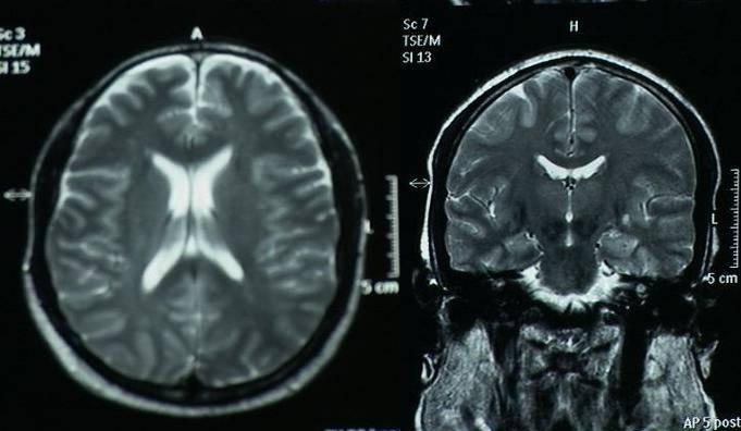
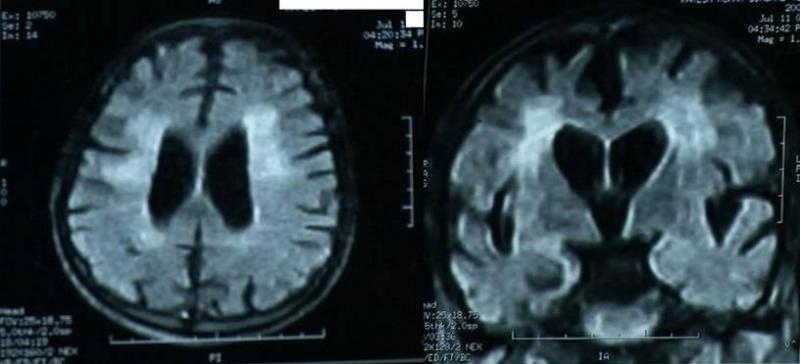
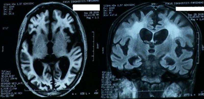
- Progressive cerebral atrophy after anoxic encephalopathy following cardiac arrest: a serial MRI study
- Mind-body interventions
- Carpel Tunnel Syndrome
- Normal pressure hydrocephalus
- Developmental delay and regression
- Brain our supercomputer
- Second opinion
- Sleep apnea syndrome
- Bumps to the head: minor concussion and post concussive symptoms
- Concussion and return to play
- Post concussive syndrome
- Coma
- Alcohol and neurodegeneration
- Alcoholic neuropathy
- Neurology of aging
- Confused mass of protoplasm
- Multiple consultations versus medical shopping
- Dementia and Aging
- Behavioral problems in dementia
- Cardiac death and organ transplantation
- Syncope
- Irritable bowel syndrome and brain
- Chronic traumatic encephalopathy
- Dr. Google
- Forgetting to learn
- Ginkgo biloba and memory
- Cyberchondria
- Statins and cognition
- Q&As
- I had a stroke like episode
- Digitalization of medical records: pearls and perils
- Brain Care Foundation
- Hypothermia and Brain Arrest Protocol
- A Doctor’s Point of View on the Doctor Patient Relationship
- Post traumatic epilepsy or seizures after head trauma
- Post traumatic epilepsy
- Incidentally discovered aneurysms in the brain-what to do about them?
- Syncope Vs Seizure: the quest for an answer
- When a seizure is not a seizure (pseudoseizures)
- Pseudoseizures
- When hospitals fail
- Disclaimer
- Contact Me
Progressive cerebral atrophy after anoxic encephalopathy following cardiac arrest: a serial MRI study